Friday, April 19, 2024
   
Text Size

Radiology

The Department of Radiology consists of highly qualified, dedicated team of radiologists working along with residents headed by Dr. Vishnuvardan Reddy. The department provides specialized, full-service which strives to meet all patient and clinician needs in diagnostic imaging and image-guided therapies. It provides both diagnostic and interventional services from computed radiography and cross sectional imaging.

Facilities:

The Department of Radiology has outstanding state of the art facilities like Magnetic Resonance Imaging (MRI), CT Scan, Ultrasound color doppler and Radiography. Well trained technical, teaching and non-teaching staff to cater all kind of radiology requirements. The classrooms and seminar halls are well equipped with LCD projectors and latest audio-visual aids. The Central Library having excellent collection of books and national & international journals on latest resource in Radiology.


Ultrasound Scan

Ultrasound (or Sonography) uses high-frequency sound waves to create an image. This technique does not involve ionizing radiation. Since images are obtained real time, our radiologists are able to observe motion and consequently assess function as well as anatomy. Common examinations include evaluation of blood vessels, gallbladder, kidneys, liver, spleen, pancreas, uterus and ovaries, urinary bladder, thyroid, fetus, musculoskeletal structures, and heart.

Doppler Ultrasonography

    A duplex ultrasound is a test to see how blood moves through your arteries and veins. The test combines traditional ultrasound with Doppler ultrasonography. Regular ultrasound uses sound waves that bounce of blood vessels to create pictures. Doppler looks at how sound waves reflect off moving objects, such as blood. There are different types of duplex ultrasound exams.
    Some include:
  • Carotid duplex ultrasound looks at the carotid and vertebral arteries in the neck.
  • Arterial and venous duplex ultrasound of the extremities to looks at vessels in the arms or legs.
  • Renal duplex ultrasound examines the kidney blood flow
  • Arterial and venous duplex ultrasound of the abdomen examines blood vessels and blood flow in the abdominal area
   

MRI

Magnetic Resonance Imaging (MRI) – is a highly advanced, painless, non invasive test that creates exceptionally clear images of the internal structures and organs of your body. It is primarily used in medical imaging to demonstrate pathological or other physiological alterations for the living tissues.

At SIMS & RH we have open MR equipment which gives us optimal diagnostic capabilities. Among out areas of expertise are neuro radiology (head, neck and spine) MRI, musculoskeletal (muscles and bones) MRI, pelvic and abdominal MRI.


X-Ray Facility

The MRL X-ray Facility provides state-of-the-art x-ray diffraction tools for characterizing structural properties of a wide range of materials including metals and composites, polymers and biological materials, and electronic and optoelectronic materials in the lengthscale range of 0.1 nm - 100 nm. As a key research asset of the MRL, the x-ray facility is accessible by MRL researchers as well as off-campus academic groups and industrial users. In addition to maintaining an array of commercial and custom-built specialty instruments, the facility staff also conducts active research in developing new x-ray imaging methods and instrumentation to advance the capabilities of x-ray characterization techniques.


CT Scan

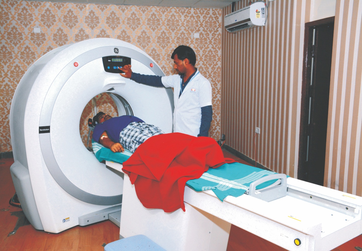
Computerised Tomography (CT) also commonly known as “CAT Scan” (Computerised Axial Tomography) provides clear three-dimensional images of body organs and their structure. These can be seen much more clearly than with conventional X-rays. The CT scans can help doctors to make an early and accurate diagnosis.

When having a CT scan you are required to lie on the CT couch which will be raised up and passed into the CT gantry. The examination involves you lying very still whilst the couch is moving in and out of the gantry. To improve the quality of images you may be required to receive an injection of contrast. This contrast is an iodine based non-ionic contrast that may make you feel warm for a short period. The contrast is used to demonstrate the arterial and venous anatomy of the area of interest.

At SIMS & RH all routine CT scans are done with single slice Siemens Esprit and 16 slice GE Light speed.

GE Light speed 16 is a state of art multi decetor CT (MDCT) scanner which generates 32 slice in 1 second. As result of the fast scanning & high tube capacity, very thin sections (0.625 mm thickness) of high resolution can be obtained in very short scanning time. CT coronary angiography, for scanning of coronary arteries is non-invasive in manner, it is possible with this scanner with high accuracy rates. Other CT angiography studies of brain, neck, abdomen, upper & lower limbs with excellent resolution comparable to digital subtraction angiography (DSA) & 3D reconstructions are also easily performed. High resolution musculoskeletal 3D CT studies. Virtual bronchoscopy, virtual colonoscopy multiphasic abdominal CT & Ct perfusion studies can also be done routinely. The routine CT studies can be done with very high resolution in very short scanning time, making it ideal for very sick patients, polytrauma patients, elderly and infants or young children. The data can be transmitted in CD also for digital storage and archiving. Patients pay the same charges for routine CT scans as those at any other centre not having these high quality machines.

   
Hackguard Security Enabled
style=][0]; ?